VNJ Articlesclinicalocularrabbits
23 August 2022
A look at ocular conditions in rabbits by Sally Turner
ABSTRACT: As pet rabbits are becoming more and more popular, they are being presented to general practice more frequently, and unfortunately eye disorders are one of the most common problems from which they can suffer. In this article, we shall consider all these lacrimating lagomorphs and blind bunnies, and try to make sense of the reasons that they can develop ocular conditions and – just as importantly – what we can do about them!
Anatomy refresher
The eyes of rabbits have the same basic anatomy as we see in all mammals – a cornea, lens, retina, optic nerve and so on. However, they have a couple of differences from dogs and cats.
Firstly they only have one nasolacrimal opening through which they can drain tears – unlike in dogs and cats which have two nasolacrimal punctae. The single rabbit punctum is located close to the medial canthus, just inside the lower eyelid. You will see why this is important later on!
The nasolacrimal duct, which drains the tears away from the eyes to the nose, is not a nice short straight tube like it is in most dogs. Instead, it twists and turns around the tooth roots of the rabbit within the maxilla and changes in diameter along its route (Figure 1). This can cause a lot of problems if it becomes blocked (which happens easily).

Figure 1: Dorsoventral view showing route of duct, (c) the nasal opening of the duct (arrow). (Reprinted with permission from pi 18, Turner, S. (2008) Saunders Solutions in Veterinary Practice Small Animal Ophthalmology Elsevier)
Another anatomical difference lies in the arrangement of blood vessels in the retina – in dogs and cats the three or four main arterioles and venules branch out from the optic disc (called a holangiotic pattern). In rabbits, there are just two main vessels which branch out horizontally from the elongated optic disc (called a merantiotic pattern).
One final important difference lies behind the globe. There is a large plexus of blood vessels present in the rabbit globe. Thus we need to be aware of this when enucleating rabbits – accidentally rupturing this mass of blood vessels can result in extreme blood loss and even death – so clearly it is very important to be aware of it!
Common ocular complaints in rabbits
Conjunctivitis
Conjunctivitis (inflammation of the conjunctiva) is a frequent complaint. One or both eyes can be affected. We see conjunctival hyperaemia, chemosis (swelling of the conjunctiva) and ocular discharge which can be serous (watery) or mucopurulent.
Irritation from dusty hay, poor hygiene with a build-up of excreta, foreign bodies from bedding and bacterial infections – staphyloccal, Pasteurella or Escherichia coli spp. – are all possible causes.
Gently bathe the affected eye(s) with sterile saline, together with appropriate antibiotic cover – (both fusidic acid (Fucithalmic – Dechra) and gentamycin (Tiacil – Virbac) are licensed for use in rabbits.
A quite unusual, but very interesting, condition we see in rabbits is that of aberrant conjunctival overgrowth, or pseudopterygium. This is a painless, often bilateral, condition in which a fold of conjunctiva grows across the cornea from the limbus, moving centrally (Figure 2). It can compromise vision if bilateral.

Figure 2: Aberrant conjunctival overgrowth – the membrane of conjunctiva has grown over the cornea from the limbus
The underlying aetiology is unknown. The membrane does not attach to the cornea and can easily be surgically excised, but usually grows back unless the remnant at the sclera is sutured directly to this to stop it reforming.
Corneal ulcers
Traumatic ulcers are not uncommon, usually caused by inappropriate bedding or fighting with other hutch-mates. Treatment tends to be the same as for dogs and cats.
First check that there is no foreign body remaining in the conjunctival sac, including under the nictitans membrane (third eyelid), then treat with a broad spectrum antibiotic topically and the ulcer should heal in a few days.
Dacyrocystitis
Dacryocystitis (inflammation of the nasolacrimal duct) is a real problem in rabbits. It can follow on from an untreated or mis-treated conjunctival infection, and also commonly is seen in rabbits with dental disease.
Here the tooth roots impinge on the long tortuous nasolacrimal duct and obstruct it – either in the region of the molars or at the incisor roots. Thus if a patient is suspected of suffering from dacryocystitis, it is very important to do a thorough dental examination, including radiography, if indicated.
Affected individuals have a sticky purulent ocular discharge, which often spills down the face and can make the periorbital skin very sore (Figure 3).

Figure 3: Dacryocystitis showing the copious discharge at the medial canthus
Some will also have upper respiratory signs (‘snuffles’). Swabs should be taken for culture and sensitivity testing, since bacteria such as E. coli and Pasteurella multocida are frequent isolates.
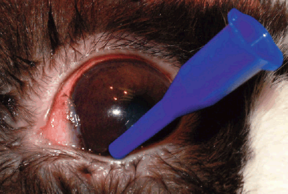
Along with frequent bathing, topical and systemic antibiotics, such as enrofloxacin (Baytril – Bayer), licensed for use in rabbits, can be administered. It is important to flush the nasolacrimal duct. This can be done in most co-operative rabbits with just topical anaesthesia.
The single, lower, nasolacrimal punctum is located and cannulated; and using a 5ml syringe, filled with sterile saline, gentle pressure is used to try to flush the discharge down the duct (Figure 4). Successful draining will result in drops of discharge appearing at the nostril.

Figure 4: Cannulation of the nasolacrimal duct to allow flushing
The flush should be repeated until clear saline is seen at the nose.
Unfortunately, some rabbits will have blocked ducts and it will not be possible to flush successfully. Antibacterial treatment should be instigated and attempts made at flushing the duct every few days.
Sometimes blockages can be cleared in this way, but some are permanent – especially in rabbits with dental disease. In these cases, the condition can be controlled by cleaning several times daily and intermittent use of systemic and topical antibiotics. In some patients, the condition becomes so severe that euthanasia is required.
Uveitis and Cataract
Uveitis (inflammation of the iris, c
iliary body and choroids) can occur following simple trauma and responds to anti inflammatory treatment. However, the microsporidian parasite, Encephalitozoon cuniculi, can sometimes be implicated. Most rabbits are clinically well with just an eye problem, but systemic involvement can occur, with neurological and renal symptoms most frequent.
The owner might notice that the eye has been inflamed, but sometimes will just notice a white lesion inside the eye (Figure 5). This usually represents an abscess from the iris or ruptured lens, and cataracts are often present. One or both eyes can be affected. Antiparasitic treatment with fenbendazole (for example, Panacur – Intervet Schering Plough, recently licensed for rabbits) is recommended along with topical anti inflammatory drops.

Figure 5: Cataract and uveitis in a rabbit with Ecuniculi infection
If the condition occurs bilaterally, with cataracts, such that the rabbit is blind, surgery can be considered. Phacoemulsication to remove the cataract can be quite successful in such cases.
Cataracts can also develop without any predisposing factors. New Zealand white rabbits have been reported with presumed inherited cataracts, and congenital ones are occasionally encountered in litters. Diabetes mellitus can occur in rabbits and cataracts may develop secondary to these, just as in dogs.
Retrobulbar disease
Exophthalmos (protrusion of the eye) is another frustrating condition which is not uncommon in rabbits. Sometimes the presentation is acute, but more often it develops gradually over a few weeks and the eye appears to bulge forwards. It can be confused with glaucoma, where the eye becomes enlarged and thus bulges.
Looking at the rabbit from above will help to differentiate the exophthlamic eye- where the normal-sized globe is pushed forwards – from the glaucomatous one, where the whole eye is enlarged.
Dental disease often accompanies ocular problems in cases of exophthalmos and patients can be inappetent and thin. Since rabbit abscesses tend to be poorly responsive to lancing and drainage, surgical removal of the abscess is advised, but this will entail enucleation (remembering about that retrobulbar vascular plexus). Unfortunately, euthanasia on humane grounds is often required.


Summary
This short article illustrates some of the similarities and differences which occur with ocular conditions in rabbits, compared to dogs and cats.
A logical approach to the diagnosis, along with careful nursing and paying particular attention to the special requirements of lagomorphs, can make treating eye conditions in rabbits, very rewarding in the majority of cases.
Author
Sally Turner
MA VetMB DVOphthal MRCVS
Sally Turner is a graduate of Cambridge University, who after a short spell in general practice carried out her ophthalmology training as a resident at the Animal Health Trust. Currently, in addition to her clinical work in private referral practice in London, Sally regularly lectures to vets and nurses and has written two books on veterinary ophthalmology (one specifically for nurses!).
Selected reading
GELATT, K. N. (2007) (Ed) Veterinary Ophthalmology. 4th Edition, Blackwell.
HARCOURT-BROWN, F. M., HOLLOWAY, H. K. R. (2003) Encephalitozoon cuniculi in pet rabbits Veterinary Record 1 52: 427-431.
TURNER, S. (2005) Veterinary Ophthalmology – A Manual for Nurses and Technicians. Elsevier Turner, S. (2008) Saunders Solutions in Veterinary Practice Small Animal Ophthalmology. Elsevier WOLFER, J. et al., (1993) in the rabbit. Prog Vet & Comp Ophthal 3(3): 91-97.
Veterinary Nursing Journal • VOL 25 • No12 • December 2010 •
