
VNJ Articlescatclinicaldogsystem
23 August 2022
Reproductive system of the dog and cat Part 1 -the female system by Victoria Aspinall
ABSTRACT: The female reproductive system has evolved to produce ova from the ovaries which are released and pass down the reproductive tract where they may be fertilised by sperm from the male animal. The parts of the tract provide a receptacle in which the resulting embryos and, later, foetuses can receive nutrition, develop and grow, and a channel along which the foetuses can pass during parturition.
The process of reproduction depends on an interrelationship between the hormones, oestrogen and progesterone and these are secreted by the ovaries. The anatomy of the bitch and queen are similar and any significant differences are highlighted.
Introduction
The female dog (Cants familiaris) is called a bilch and the female cat (Felis cat us) is called a queen. The reproductive tracts of both species are very similar and vary mainly in size. Both species are litter-bearing or multiparous, so the tract is designed to carry many foetuses in a single pregnancy.
The shape of the uterus is bicornuate – with two long horns and a small body (Figure I) The fertilised ova implant at equal distances along the horns allowing the maximum amount of space for development – it is rare for a foetus to develop within the uterine body.
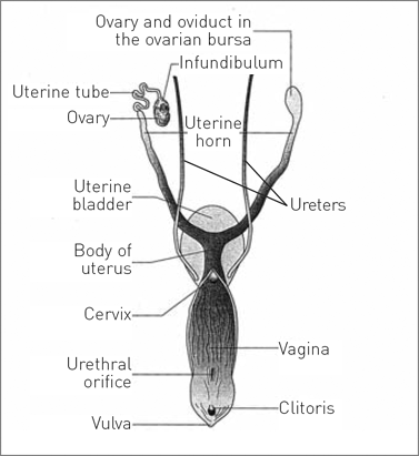
Figure 1: Dorsal view of the reproductive system of the bitch. [Reproduced with permission from Aspinall, V. and Capello, M. (2009) p128.)
Functions
The functions of the female reproductive tract are to:
• release ova to be fertilised by the spermatozoa of the male
• provide a receptacle for the development of the foetuses
• secrete the hormones oestrogen, from the developing follicles and progesterone, from the corpus luteum. These, by differing mechanisms, ensure the correct environment for the production of ova, for the survival of sperm and ova and for the development of the fertilised ova into viable foetuses.
In the non-pregnant female, most of the tract lies within the abdominal cavity, although the cervix, vagina and vestibule lie in the pelvic cavity. During pregnancy the weight of the foetuses pulls the cervix over the pelvic brim into the abdomen.
Reproductive tract
Ovary
There is a pair of ovaries – one lying on either side of the midline, caudal to each kidney – in the dorsal abdominal cavity. Each ovary is held close to the kidney by the ovarian ligament, which contains smooth muscle fibres allowing it to stretch in response to the weight of the pregnant uterus.
Each ovary is suspended from the abdominal wall by the mesovarium which is part of the visceral peritoneum. Part of the mesovarium forms a pocket¬like ovarian bursa in which there is a small opening – this is the only means of entry into the peritoneal cavity.
The ovarian tissue consists of a network of connective tissue and blood capillaries, derived from the ovarian artery. Within this are germ cells that eventually develop into follicles, each containing an ovum.
The function of the ovary is to produce ova and to secrete oestrogen and progesterone.
Uterine tube
This is also called the oviduct (or the Fallopian tube) and its function is to conduct the ova from the ovary to the uterine horn. Each tube is a narrow structure lying close to – and running over – the ovary and leading into the uterine horn.
The open end, close to the ovary, is a funnel-shaped infundibulum which is fringed with finger-like fimbriae. The infundibulum is able to move over the surface of the ovary to capture an ovum as it ovulates from its follicle. The tube is suspended by the mesosalpinx which is continuous with the mesovarium.
Internally the tube is lined with ciliated columnar epithelium. The hair-like cilia assist in the movement of the ova down the tube and the lining membrane secretes nutrients which aid the survival of the ova as they pass down and of the spermatozoa as they pass upwards. Fertilisation occurs in the uterine tube.
Uterus
The uterus is a central Y-shaped structure consisting of two long uterine horns and a small uterine body (Figure 1). Its function is to provide a receptacle and the correct environment for the development of the fertilised embryos into foetuses, and also to provide the means for the development of a placenta which supplies nutrients to the developing conceptus.
The uterus is held in position within the abdominal cavity by the broad ligament or the mesometrium, which is continuous with the mesovarium and mesosalpinx. Running within the mesometrium are the uterine and ovarian arteries and veins that supply the tract with blood – the uterine artery runs close to the cervix and is ligated during a bitch spay.
The wall of the uterus consists of three layers:
1. endometrium – columnar epithelium, glandular tissue and blood capillaries. This layer enlarges during pregnancy to enable implantation of the placenta.
2. myometrium – smooth muscle fibres which contract strongly during parturition to expel the foetuses.
3. mesometrium – part of the visceral peritoneum.
Cervix
The cervix connects the uterine body with the vagina and is a thick-walled muscular sphincter through which runs the narrow cervical canal. The canal is usually closed but it relaxes to allow the passage of sperm or foetuses. During pregnancy, the canal is blocked by a mucoid plug which protects the conceptuses from infection.
Vagina
This structure is a dilatable tube which extends from the cervix to the external urethral orifice (Figure 1). It lies within the pelvic cavity and is surrounded by connective tissue. Internally it consists of longitudinal folds which dilate to allow the foetuses out during parturition.
The vagina is lined with stratified squamous epithelium, which changes in response to the hormones of the oestrous cycle. In the bitch, daily vaginal smears may be examined to monitor the progress of the cycle prior to mating – this is known as exfoliative vaginal cytology. During pro-oestrus, the blood-stained discharge comes from the vaginal lining, not from the uterus, as occurs in man.
Vestibule
The vestibule is a continuation of the vagina (Figure 1) running from the externa] urethral orifice – the point at which the urethra leading from the bladder joins the tract – to the outside at the vulva. Its function is to convey foetuses out of the body and sperm into the vagina from the male penis and also to convey urine from the bladder and urethra out of the body. Its structure is similar to that of the vagina, but the walls are not ridged by longitudinal folds.
Vulva
The vulva marks the external opening of the tract and lies in the perineum, ventral to the anus, below the tail. It consists of two vertical labiae joined dorsally and ventrally with the vulval cleft between them. Just inside the ventral part of the cleft is the clitoris made of cavernous erectile tissue. This is the equivalent of the male penis.
Normally the labiae are held tightly together to prevent the entry of infecti
on, but in the bitch, during pro-oestrus and oestrus, the vulva enlarges and is slightly relaxed. This does not happen in the queen.
Mammary glands
The dog and the cat are members of the class Mammalia and the defining characteristic of this group is the presence of mammary glands, which secrete milk on which the young are fed. Mammary glands are theoretically part of the integument – as opposed to the reproductive tract itself – as they are modified skin glands.
In the bitch and the queen, they lie externally on the ventral abdominal and thoracic body walls, just below the skin on either side of the midline. The bitch has five pairs, while the queen has four pairs. Mammary glands are present in both sexes.
Each mammary gland consists of glandular tissue embedded in connective tissue and lined by a secretory epithelium (Figure 2). The milk, produced during a process known as lactation, drains through a network of sinuses that eventually narrow to form teat canals. Each teat canal leads to a teat orifice on the surface of the teat – there are many orifices on one teat so the milk sprays out into the neonate’s mouth.
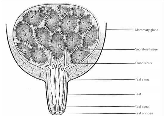
Figure 2: Breeding from a mare is uniquely satisfying, especially when there is a successful outcome
Lactation is influenced by three reproductive hormones:
• progesterone – secreted by the corpus luteum within the ovary and causes the mammary glands to enlarge during the oestrous cycle and during pregnancy.
• prolactin – secreted by the anterior pituitary glands and causes the glands to secrete milk during the last third of pregnancy, but at this stage it is not released.
• oxytocin – secreted by the posterior pituitary gland during the few hours around parturition. As the neonate begins to suckle the sensation starts a reflex arc which releases oxytocin which in turn causes contraction of the smooth muscle around the mammary glands and the milk is released.
Author
Victoria Aspinall bvsc mrcvs

Victoria qualified from Bristol University vet school and went into small animal practice. After raising her four children she went into teaching at Hartpury College, where she started the veterinary nursing department. From there she founded Abbeydale Vetlink Veterinary Training which is a VNAC in the west of England and also runs a wide range of veterinary-related courses, including vet nurse training.
Victoria is now an associate lecturer in veterinary nursing at Bridgwater College, Somerset. She has written and edited many books for vet nurses, including The Complete Textbook of Veterinary Nursing.
To cite this article use either
DOI: 10.1111/j.2045-06il8.2010.00013.x or Veterinary Nursing Journal Vol 26 pp 43-45
Bibliography
ASPINALL. V. and CAPELLO, M (2009) Introduction to Veterinary Anatomy and Physiology 2nd ed. Butterworth Hememann, Oxford DYCE. K M. SACK. W 0 and WENSING. C. J. G 120021 Textbook ol Veterinary Anatomy 3rd ed. Saunders.. Philadelphia.
EVANS, H. E. 119931 Milter s Anatomy of the Dog 3rd ed. Saunders. Philadelphia
Veterinary Nursing Journal • Vol 26 • February 2011 •