VNJ Articlesclinicalreproductivesystem
23 August 2022
Reproductive system of the dog and cat Part 2 -the male system by Victoria Aspinall
ABSTRACT: The male reproductive system has evolved to produce spermatozoa by a process of spermatogenesis, to secrete fluids which both aid the survival of the sperm and transports them into the female tract, and also to secrete the hormone testosterone which is essential in the development of male characteristics and patterns of behaviour. The parts of the reproductive trad all play a part in these functions and in delivery of the sperm into the female tract, where they are able to fertilise the female ova.
The anatomy of the reproductive systems in the dog and the tomcat are similar and any significant differences are highlighted
Introduction
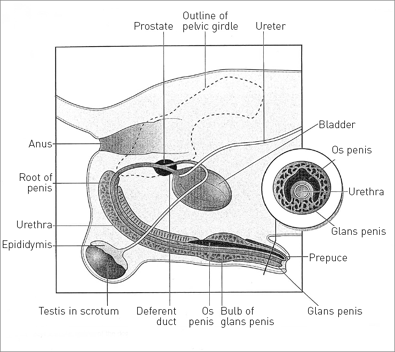
The male canid is known as a dog and the male felid is known as a tomcat. The reproductive tract of both species is similar, but there are some significant differences that will be highlighted later in the article (Figures 1 & 2).

Figure 1: Reproductive system of the dog. Images are reproduced with kind permission of Victoria Aspinall and Melanie Capello

Figure 2: Reproductive system of the tomcat. Images are reproduced with kind permission of Victoria Aspinall and Melanie Capello
Function and anatomy
The functions of the male reproductive system are to:
• produce spermatozoa (sperm) by a process of spermatogenesis, which will fertilise the ova produced by the female
• secrete fluids which aid the survival of the sperm and transports them into the female tract during coitus
• secrete the hormone testosterone, which influences the development of secondary sexual characteristics and male behaviour patterns.
Testis
There is a pair of testes the main function of which is the production of sperm (spermatogenesis) which only occurs below temperatures of 40°C. In the adult, the testes lie outside the body cavity within a pouch of pigmented and sparsely furred skin called the scrotum (Figure 3). In the dog this lies between the upper thighs, whereas in the cat it lies ventral to the anus, close to the ischial arch.

Figure 3: A. Testis within the scrotum; B. Scrotum removed; C. Cross-section through the seminiferous tubules. Images are reproduced with kind permission of Victoria Aspinall and Melanie Capello
Within the skin is the Dartos muscle, which in cold weather contracts and pulls the testes closer to the warmth of the body; in warm weather, it relaxes and the scrotum and testes drop away from the body allowing them to remain cool.
The majority of the testicular tissue consists of coiled seminiferous tubules lined by spermatogenic cells responsible for the formation of sperm, and by Sertoli cells which secrete nutrients to prolong the life of the sperm and small quantities of the hormone, oestrogen. Lying between the seminiferous tubules are the cells of Leydig or interstitial cells that secrete testosterone.
The coiled seminiferous tubules eventually unite to form the epididymis. This is a long, coiled tube lying within a sheath along the dorso-lateral border of the testis. It becomes the cauda epididymis at the caudal extremity of the testis and at this point the temperature is at its lowest. Here the sperm, formed in the tubules, undergo a process of final maturation and are stored ready to be propelled into the remainder of the male system and into the female tract during ejaculation. The epididymis continues as the deferent duct which passes out of the scrotum into the peritoneal cavity via the inguinal ring.
Within the scrotum, the testis is wrapped in an evagination of the peritoneum forming a double layer of tissue known as the tunica vaginalis (Figure 3). This also wraps around the spermatic cord which contains the testicular artery, vein and nerve and the deferent duct. As it enters the scrotum, the testicular artery forms a complex network of arterioles called the pampiniform plexus which ensures that the blood is cooled before it enters the testicular tissue.
Within the spermatic cord is the cremaster muscle. This works in conjunction with the Dartos muscle to raise or lower the scrotal sac to adjust the temperature to facilitate spermatogenesis.
Deferent duct
The deferent duct is known as the vas deferens or the ductus deferens, and is a continuation of the cauda epididymis running within the spermatic cord. It enters the peritoneal cavity through the inguinal ring, which is a split in the aponeurosis of the external abdominal oblique muscle in the groin of the animal.
The deferent duct from each testis joins the urethra at a point within the prostate gland (Figures 1 &2) and here the walls of the two ducts are thickened and glandular.
Urethra
From the point at which the deferent ducts join the urethra, the tract is shared by both the urinary and reproductive systems. The urethra runs from the neck of the bladder to its external opening at the tip of the penis. In the dog, the urethra is long and has a pelvic part running along the floor of the pelvis and a penile part running through the body of the penis (Figure 1) – its opening pointing cranially.
In the tomcat, there is a short preprostatic urethra between the neck of the bladder and the prostate gland (Figure 2). The penile urethra is short and does not extend further than the ischial arch – its opening points caudally and is located ventral to the anus. This position is related to the tomcat’s habit of scent marking its territory by spraying vertical surfaces with urine at the nose height of other cats.
Accessory glands
The tract is supplied by the prostate gland present in the dog and the cat and lying close to the neck of the bladder (Figures 1 & 2) and the bulbo-urethral glands – present only in the cat. These lie close to the tip of the penis.
The function of both types of glands is to produce secretions which increase the volume of the ejaculate, helping to propel it into the female tract during coitus. The secretions have an alkaline pH which neutralises the acidity of any urine within the urethra and provides the correct environment for sperm survival.
Penis
As the urethra leaves the pelvic cavity, passing over the ischial arch, it becomes surrounded by cavernous erectile tissue forming the corpus cavernosum penis.
This tissue consists of‘caverns’ of connective tissue lined with endothelium which, during sexual excitement, become tilled with blood under pressure resulting in engorgement of the organ allowing it to be introduced into the female vagina during coitus.
There are significant differences between the anatomy of the penis in the dog and the cat.
Dog
The dog’s penis, consisting of the central urethra and the surrounding corpus cavernosum penis, is attached to the ischial arch by a pair of muscular crura, which come together in the midline to form the root of the penis.
It then curves cranioventrally between the upper thighs of the dog, forming the body of the penis attached to the ventral body wall and the glans penis which is its free end and makes up about a quarter of the length.
Within the tissue of the glans penis – and lying dorsal to the urethra – is a small bone, the os penis. During the early stages of coitus, erection is not complete until the vaginal muscles of the bitch clamp around the penis; so the function of the os penis is to aid rigidity and entry into the vagina at this early stage. The urethra runs through the bony tunnel of the os penis and as it is unable to expand at this point it may be a site for blockage with urethral calculi.
Tomcat
In the tomcat, the length of the urethra surrounded by cavernous erectile tissue is short and at this point there is the opening of the bulbo-urethral glands. Within the erectile tissue ventral to the urethra is a bony os penis. Within this area the urethra is narrow' and it is here that blockage by struvite crystals is most likely to occur.
The glans penis is covered with tiny backward-pointing barbs. After coitus, as the tomcat withdraws his penis from the vagina of the queen, these barbs cause a moment of intense pain – the queen will often give a loud howl. This pain initiates a reflex arc, involving the pituitary gland and the ovary, and resulting in ovulation 36 hours later – the queen is an induced ovulator.
During mating the penis engorges and curves cranioventrally so the mating position is similar to that seen in other mammals.
Prepuce
When relaxed, the entire length of the penis of the dog and cat lies inside a protective prepuce. The outside is covered in hairy skin and the lining consists of a mucous membrane which is continuous with that of the urethra and is well supplied with lubricating glands. Infection of the prepuce is known as balanoprosthitis and results in an unpleasant greenish discharge.
Author
Victoria Aspinall bvsc mrcvs

Victoria qualified from Bristol University vet school and went into small animal practice. After raising her four children she taught at Hartpury College, where she started the veterinary nursing department. She subsequently founded Abbeydale Vetlink Veterinary Training which is a VNAC in the west of England.
Victoria is an associate lecturer in veterinary nursing at Bridgwater College, Somerset. She has written and edited many books for vet nurses.
To cite this article use either
DOI: 10.1111/j.2045-0&48.2010.00025.X or Veterinary Nursing Journal Vol 26 pp89-91
Bibliography
ASPINALL, V. and CAPELLO, M, 120091 Introduction to Veterinary Anatomy and Physiology 2nd ed. Butterworth Heinemann. Oxford.
DYCE, K. M., SACK, W. 0. and WENSING. C. J. G. 120021 Textbook of Veterinary Anatomy 3rd ed Saunders. Philadelphia.
EVANS. H. E. 119931 Miller's Anatomy of the Dog 3rd ed- Saunders. Philadelphia.
Veterinary Nursing Journal • VOL 26 • March 2011 •
