ABSTRACT: Vision is an importanl sense, not only for people, but also for our dogs. Ocular health, therefore, is an important aspect of veterinary medicine.This article gives an overview of the ocular lens anatomy and physiology. It also explains the two most common diseases of the lens: cataracts and lens luxation. For both conditions, pathogenesis, as well as subsequent changes within the eye, is described.
The lens is a highly specialised tissue that is responsible for focusing sharp images onto the retina. To fulfil its function, it has to be transparent and requires a refractive surface. In most species, the lens is also able to change its refractive power to allow focusing objects that are seen at different distances (accommodation).
Anatomy and physiology
The lens is positioned in the anterior part of the eye, just behind the iris (Figure 1). It is kept in place by inelastic microfibrils, named zonules. The zonules insert near the lens equator at 360° and attach the lens to the ciliary body. The lens consists of an outer lens capsule and onion-like concentric layers of lens fibres forming a central nucleus and peripheral cortex (Figure 2). The lens fibres are constantly produced throughout life.

Figure 1: Schematic picture of the globe

Figure 2: Schematic picture of the lens anatomy
The lens lacks vascularisation. It therefore depends on the surrounding fluid, the aqueous humour and, to a lesser extent, the vitreous (hydrogel in the posterior segment of the globe) for its metabolic needs. Hence it is sensitive to changes in either of these compartments, which commonly results in cataract formation.
Cataracts
Cataracts are a leading cause of visual impairment in dogs. They are opacities of the lens caused by changes of the lens protein or hydration (Figure 3). While focal cataracts may have a limited effect on vision, more advanced cataracts can result in blindness, significantly impairing the quality of life in our patients. Cataracts can be congenital or acquired.

Figure 3: Nine-year-old cross-bred dog with a total cataract in the right eye
Congenital cataracts are present at birth and frequently associated with other malformations, such as microphthalmus (abnormally small eye), but can present as a sole abnormality. They affect the central aspect of the lens (nucleus) which develops earliest in life. Nuclear cataracts are often non-progressive and surrounded by newly formed, transparent lens fibres. They can be inherited or a consequence of an insult in utero.
Acquired cataracts can be the consequence of an hereditary defect. These cataracts can occur in very young animals or might become apparent later in life. Hereditary cataracts usually follow a certain pattern within a specific breed and are comparable with regards to their location within the lens and age of onset. The mode of inheritance of cataracts is only known for a small number of breeds and genetic tests for cataract-causing mutations are available – at the Animal Health Trust – for the Australian shepherd dog, Boston terrier, French bulldog and the Staffordshire bull terrier.
Causes for secondary cataracts include ocular diseases, such as uveitis (intraocular inflammation) or progressive retinal atrophy, as well as systemic diseases including diabetes mellitus. External sources, such as trauma, UV light, radiation and toxins have also been associated with cataract development.
Cataracts and diabetes mellitus
Diabetes mellitus is a very common cause for cataracts. Owing to the increased amount of glucose in the lens and aqueous, the metabolism within the lens changes resulting in uptake of fluid into the lens and subsequent loss of lens transparency.
One study shows that half of all diabetic dogs will develop cataracts within 170 days after diagnosis, while 75 per cent and 80 per cent of dogs will be affected by 370 and 470 days respectively. Therefore, owners of diabetic patients should be prepared early for this common complication.
Diabetic cataracts often progress very rapidly and the swelling of the lens can result in a rupture of the lens capsule and subsequent violent lens-induced uveitis.
The latter can result in sight-threatening complications, such as retinal detachment and glaucoma. Hence diabetic patients with sudden onset cataracts should be assessed early for possible cataract surgery, even when the diabetes mellitus is not yet fully stabilised.
Redness of the eye, a bluish appearance of the cornea (corneal oedema), an irregular pupil and pigment dispersion on the lens are suggestive of lens induced uveitis and should prompt emergency referral.
Lens luxation
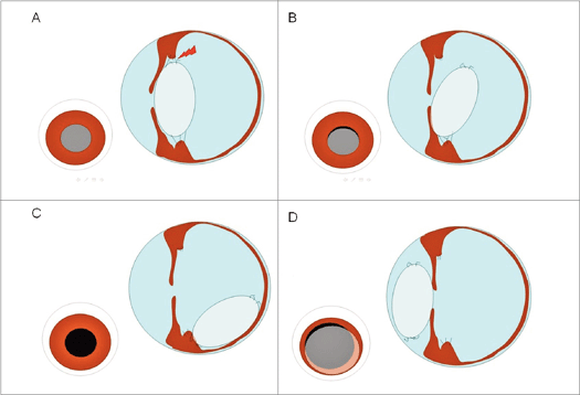
In cases of lens luxation, the zonules that keep the lens in place lose their strength (Figure 4). A lens luxation can be primary, caused by a genetic defect in the zonula anatomy. This has been found to be an inherited condition in many terriers (Jack Russell terrier, Parson Russell terrier, Wire-haired Fox terrier, Miniature Bull terrier, Tibetan terrier, for example) but also in some other dog breeds (Lancashire Heeler, Border collie, for instance).

Figure 4: Damage to the zonules (A) will result in subluxation IB], posterior (C] or anterior ID) luxation of the lens
In the affected dog, the zonules start to break down during their early life, resulting in increased movement of the lens. Subsequently, the lens ‘disinserts’ from the surrounding tissue and initially subluxates (partial disinsertion) – eventually luxating completely. As a consequence, the moving lens damages other intraocular tissues which may lead to blindness.
A secondary lens luxation occurs when other conditions cause the zonula defects. An ocular trauma or glaucoma (increased intraocular pressure) with subsequent enlargement of the eye, can result in stretching and eventually rupturing of the zonules. In cases of uveitis, the inflammatory response can also have a destructive effect on the zonules.
Notably, anterior lens luxation (luxation of the lens in the a
nterior chamber of the eye) can result in very acute blindness (Figure 5). The lens impairs the normal flow of fluid (aqueous) inside the eye which leads to a rapid increase of the intraocular pressure with subsequent damage to other ocular tissues. The retina in particular, where light energy is converted into nerve impulses, is sensitive to increased intraocular pressure. Its damage can lead to transient or permanent blindness, depending on the altitude in intraocular pressure and the time span of the increase.

Figure 5: Anterior lens luxation in the right eye of a 4 Vi -year-old Yorkshire terrier
An anterior lens luxation, therefore, always presents as an emergency, and surgical removal of the lens is usually indicated. However, movement of the lens inside the eye can also result in damage to the cornea and retina.
In an article next month, Claudia Busse will address the management of affected patients, treatment options and the nursing aspects of providing optimal care for dogs with cataracts and lens luxation.
Author
Claudia Busse DVM MRCVS

Claudia Busse graduated from the School of Veterinary Medicine, Hanover. Germany in 2004. Afterwards, she completed a doctoral thesis on inherited ocular diseases and worked in general practice. In 2005, she started a rotating internship at the Animal Health Trust in Newmarket and subsequently completed a residency in veterinary ophthalmology. Claudia currently works at the AHT as a clinician in ophthalmology.
To cite this article use either
DOI 10.1111/j.2045-0648.2010.00003.x or
Veterinary Nursing Journal Vot 26 pp12-14
Further reading
8SAVA Manual of Small Animal Ophthalmology. Eds. Simon Petersen-Jones and Sheila Crispin, 2nd edition, BSAVA Gloucester.
Veterinary Ophthalmology, Ed Kirk N Gelatt, 4th edition. Blackwell Publishing
• VOl 26 • January 2011 • Veterinary Nursing Journal
